electrocardiogram (EKG) : what to expect

HCE242_2.jpg

Of all the experiences in our clinic, the EKG (or ECG) is probably the part that you should literally have no concerns about. The EKG is one of the simplest and most routine diagnostic studies done for your heart. It is designed to monitor the electrical activity of your heart. Why is this?  Your heart's electrical activity can tell your cardiologist a lot of information about its function.

In the office, one of our medical assistants will help get you setup for this procedure. You will lie flat on one of our examination tables, with your upper body exposed, and 12 electrical wires, will be attached to your chest with small pieces of sticky tape. Don't worry, these wires don't deliver any electricity, but are rather designed to measure the electrical activity of your heart from the surface of your chest. Once you are setup, you will need to stay perfectly still, as the EKG machine carefully listens to your heart from all 12 locations.

The machine will then interpret the electrical activity that it detects and plot that on a piece of paper showing the waveform of the electrical current that it detects.

This EKG print out will then be interpreted by your cardiologist. This print out has a remarkable wealth information to the trained eye. Your cardiologist can use this information to detect information about your hearts' rhythm centers, the conduction of electricity through your heart which signals to the different chambers of your heart how to contract, whether or not you've had a heart attack, and also a number of other interesting findings such as the size of your heart chambers, whether or not your electrolytes are balanced and even whether or not you may have lung disease! It is a remarkable tool that gives an amazing breadth of information to help treat you better.

Usually after the EKG is complete, the leads and the sticky tapes all come off, and you are back to waiting for the doctor usually in a matter of minutes!

Curious? See to your left what an EKG actually looks like. Shown here is an EKG from a normal heart. Remember that just as God did not create everyone the same, no two EKGs look exactly the same. Just because your EKG does not look exactly like the one here does not mean that yours is abnormal. There is a great degree of normal human variation that doctors train for years to be able to take into account when they interpret your study.


stress testing : what to expect

A stress test can be both a confusing and daunting experience to many patients, simply because they do not know what to expect. Let's first understand what a stress test is designed to test, and then explore the things patients should know.

In our offices, stress testing is performed in collaboration with the Memorial Hermann Sugar Land Hospital. This means, that our stress tests are performed by the hospitals' expert staff who have supervised and performed thousands upon thousands of stress tests. 

Your doctor might prescribe a stress test in a myriad of different forms. Briefly, all stress tests are designed to address two questions, and they are each addressed by separate components or parts, of the stress test. First, they ask what the functional capacity of the patient is. This is called the stress component. Second, they ask whether blood flow to the heart is compromised during peak exertion (perfusion component).  If blood flow to the heart is compromised during peak exertion, that would strongly suggest that there is a plumbing problem, and that one or more blood vessels supplying blood to the heart is narrowed or completely blocked. Usually when people have blockages or narrowing of blood vessels that supply blood to the heart, this is called coronary artery disease. So the purpose of the stress test (with both stress and perfusion components) is to get a reasonably good estimate of whether you may or may not coronary artery disease. 


STRESS COMPONENT

The stress component of the stress test evaluates your functional capacity. Functional capacity simply means how much vigorous physical activity your heart can tolerate. You might wonder why your doctor cannot simply ask you this question instead of testing you? The reason for a formalized test is so that cardiologists around the world can standardize your functional capacity. This enables us to predict exactly how someone of your age should perform if they are generally in good shape. Let's take an example. Through analyzing lots of data, cardiologists know that someone who is 50 years old should be able to achieve a peak heart rate of about 170 beats per minute (bpm). If you can roughly reach 85% of that, or 145 bpm, then your heart can be expected to sufficiently pump blood without functional limitation. This is what the first part of the stress test measures.

In a majority of cases, the stress component of your study will be done with the use of a treadmill.  This is called a exercise treadmill stress test.  In this study, you will run on a treadmill for as long as you can. This is very important so let me repeat it. You should go on the treadmill for as long as you can. Don't stop because you think the study is over. You need to go until you are completely fatigued, or until you have some symptom that prevents you from continuing.When you are on the treadmill, your heart rate, blood pressure, oxygen saturation, and your heart's electrical activity will be monitored in real time. This information is being fed into a computer that processes this information to see how your heart is behaving and adapting to the increasing workload. Every 3 minutes, the treadmill will get harder (the incline will get steeper and the belt will start moving faster). When you think you are going to need to stop because you are too tired, too short of breath, having chest pain, leg pain or feeling some other symptoms, let the staff know ahead of time so that they can stop the treadmill safely.

When the treadmill comes to a complete stop, you will continue to be monitored for about another 5-10 minutes in recovery to ensure that your heart can relax after strenuous activity.

In many cases, this ends the treadmill exercise stress test. 

how to prepare for your stress test

  • Drink plenty of fluids the day before, and get a good night's rest. 
  • Your dinner on the day before your stress test should be your last meal. Do not eat or drink anything after midnight. 
  • If you have medications that you need to take at night, take them with your dinner. If you have morning medications, either postpone taking them until after the stress test or bring those medications with you.
  • Avoid anything with Caffeine! - This means soda/carbonated beverages, coffee (including decaf), tea or chocolate.
  • If you are scheduled to take a class of medications called beta blockers (this includes Metoprolol, Labetalol, Carvedilol, Bisoprolol and their trade names), please do not take the dose the evening before. If you are unsure, make sure you speak to your cardiologist or his office staff to verify. 
  • Wear light clothing and good, comfortable, running shoes like sneakers.


What HAPPENS IF I FAIL THE STRESS TEST?

The short answer is, nothing happens. It is fairly common for some people to not be able to exercise enough to get their heart to work hard enough. When this happens,  it is impossible for us to accurately assess the patients' functional capacity.  If this happens to you, don't despair. By itself, it doesn't mean much. Many patients are unable to reach an adequate heart rate or unable to exercise sufficiently during their stress component. It could be even due to completely unrelated symptoms such as feeling light headed because of low blood sugar, pain in the leg, or arthritis or joint pain due to a hurt knee. Whatever the reason is, if you told by the stress lab staff that your failed the stress test, or could not get your heart rate high enough, it just means we will need to stress you using another strategy. Let's talk about how we do that.


The lEXISCAN or "CHEMICAL" stress test

When patients are unable to exercise adequately, we can stimulate their heart to behave as it would during exercise by giving you a medication called Regadenason, or commonly known by its tradename "Lexiscan". This chemical, when injected through your IV will cause your heart to behave as it would when it is exercising. It will force the heart to enlarge its blood vessels and allow more blood to flow through them, as would be the case if you were vigorously exercising. When you get the chemical stress test, for that reason, you will feel like you are exercising although you are simply resting on the exam table. You may feel fluttering in your chest, you may feel your breath quicken, and you may notice that you are getting flushed, or sweaty. This is all to be expected. Remember though, that the chemical stress test should be given very cautiously in patients who have had lung disease (asthma, COPD, bronchospasm) or a very slow heart rate. If you have had any of these conditions, make sure you notify the staff before the chemical stress test is started.

PERFUSION COMPONENT

UPPER_LIMIT_slices.jpg

If you pass the treadmill stress test or have a chemical stress test done, your cardiologist may decide to proceed to the perfusion component. In this study, we will take pictures of your heart, and compare how the blood flow to your heart was like before and after inducing stress (via a treadmill or by giving the chemical through the IV).

In order to do the perfusion component, you will be administered a radioactive chemical tracer. Don't worry, our nuclear technologists are experts at handling these chemicals. This special chemical tracer is given through your IV. It is a radioactive substance that will enter your bloodstream and enter the coronary arteries (the blood vessels that supply blood to your heart) and selectively incorporate itself into your heart muscle. 

You will then be asked to sit or lay flat in a specialized camera called a SPECT camera which can directly see into your heart and see the radioactive tracer. It will capture images of your heart where your heart is quite literally "glowing" once the study is completed. The beautiful donut shaped images  you see on your left are images of a human heart captured by a SPECT camera. By comparing the distribution of the chemical before and after stress, we can figure out whether there is blood flow limitation into your heart with peak exercise. This is the principal behind the perfusion component of the stress test.


commonly asked questions about stress testing:

1. how long does the whole procedure take?

The stress test itself is a short procedure. But there can be a lot of waiting time before and after. For example, if you get a treadmill stress test with perfusion, you will check in at the front desk at the hospital, wait for the staff to usher you into the stress lab area, and then have the rest imaging part done, then go on the treadmill for the stress component, and then have another series of stress images done. The whole procedure can therefore take in the order of 4-6 hours. We recommend that you take a half day off, at the very least.

2. Will I RECEIVE ANY SEDATION DURING THIS PROCEDURE?

The short answer is no. Stress testing requires no procedural sedation. 

3. CAN I DRIVE MYSELF BACK HOME OR DO I NEED A SOMEONE TO GIVE ME A RIDE?

You will not need a ride back home, if you can drive. The actual test itself does not impact your ability to drive after the fact. Of course, should you have some untoward reaction to the medication or require hospitalization then you will definitely need someone to drive you back, but this is not the norm for a vast majority of our patients.

4. HOW LONG DOES IT TAKE TO GET THE RESULTS?

Your cardiologist will generally call you on the same day, or rarely the next day, once the study is read. During that phone call we will discuss with you the options (what happens next) and when you will need to follow up with us.

5. when can i eat?

You can usually eat while you are finished with the stress component and waiting. Our stress lab staff knows exactly when the timing for this is, and it is best to ask them before you eat or drink anything. If you have a critical medication that you need to take, please discuss this with our staff. They can help figure out what the best course of action is.

6. WHAT HAPPENS IF I FAIL THE TREADMILL TEST?

As noted above, it's not a big deal. If you fail the treadmill test, we have options and can administer the chemical stress test instead, provided, that you were scheduled to originally have a stress test with perfusion. In the rare times when you were only scheduled for a treadmill stress test (without the perfusion study) and you fail the stress test, you may need to be rescheduled. This has nothing to do with you, and has everything to do with the complex intricacies of insurance pre-authorization. It's not the end of the world, and we can have you rescheduled quickly for the next appointment available. Our office staff will help with this process.

7. WHAT HAPPENS IF MY COPD/ASTHMA CAUSES ME TO HAVE A BAD REACTION DURING THE CHEMICAL STRESS TEST?

This is not a problem either, because luckily, we have excellent antidotes to the chemical we give you that can be given to quickly reverse its effects. In fact, reversing the effects of the chemical has no impact on the stress test itself and we can usually proceed to the perfusion component without a hiccup.

8. what happens next?

Usually, after the stress test is complete, you go home. Your cardiologist will then have an opportunity to examine the images captured from the SPECT camera and will call you with the results. Depending on what we find, you may be asked to come for a follow up appointment in the office, or maybe scheduled for a cardiac catheterization procedure to look at the arteries of your heart more directly. 


transthoracic echocardiogram (echo) : what to expect

The transthoracic echocardiogram, commonly referred to as an "echo" is a very simple, non-invasive procedure. It is designed to allow your cardiologist to take a good look at the way your heart looks and how well it functions. The best way to think about this is to recall sonograms performed in an obstetricians' office to look at a pregnant mothers' baby. Well, an echo is exactly the same thing; except in this case the baby is your heart. The echo can not only show your cardiologist the structure and dimensions of your heart, but it can also measure complex parameters like blood flow speed, pressure drop offs etc, giving an intricate look into the function of your heart as well. This is fabulous news for cardiologists, because this gives us a mechanism to look directly into both structure and function of your heart, without even as much as a single needle stick. Isn't that amazing? 

In our office, echoes are generally performed right within the comfort of the office itself, or rarely, at an outside facility, and sometimes in the hospital. Where we choose to get the echo depends largely on the day you visit us, and the urgency with which we need to have this information. 

Echo Machine

Echo Machine

The machine we use for this purpose, called an echo machine is essentially a large computer with a probe attached to it. This probe emits ultrasound energy, which is not visible or detectable by the patient. When the probe is placed on the your chest right above the heart, the probe sends out ultrasound signals that penetrate  your skin, bone, soft tissue, muscle and reaches your heart. It penetrates the full extent of your heart, and bounces back off the heart muscle and returns back into the probe. Depending on how much energy was lost when it makes this trip and how long it takes, the computer in the echo machine can translate that information into an image, which shows us what your heart looks like.

On the left of the screen is what an echo machine looks like. The operator of the machine, also known as the sonographer will accompany you into a private room, where you will be asked to change into a hospital gown. Your chest will be exposed, and EKG leads will be attached to your chest so that the machine can not only see your heart, but also monitor your heart rate and rhythm.

The sonographer will then use a lubricant (also called "ultrasound jelly") and apply a little bit of this to your chest and place the echo probe (as shown in this picture) over your chest. When he or she does so, the computer in the echo machine will display an image of your heart in real time.

The echo provides two types of information to cardiologists. First, it shows us the structure of your heart. This includes the size of your heart muscle, the size of the heart chambers, and the presence of any unexpected growths or blood clots within your heart.

Second, the echo can also give us function information of your heart. By using complex mathematics, the echo machine can actually measure how fast blood flows through your heart, how stiff your heart muscle is and how rapidly it recoils after contracting. It can also give us an incredible amount of information about the function of your heart valves, including whether or not they are narrowed (called stenosis) or whether there those heart valves are leaking (called regurgitation). If you'd like to know more about heart valve disease, read up about this on our section on heart valve diseases

WHAT TO EXPECT BEFORE AN AFTER YOUR ECHO

The echo is a simple, non-invasive, office-based diagnostic test. In some ways, it is even less invasive than having your labs drawn (blood tests). There are no needle sticks, no anesthesia or sedation and nothing you need to do on your part in preparation for it. You will go home immediately after the test is complete.


trans-esophageal echo (TEE) : WHAT TO EXPECT

In principle, a transesophageal echocardiogram, commonly known as a "TEE"  is actually the same as a regular echocardiogram. The main difference is the location from where the images are acquired. In a regular echocardiogram, a sonographer will place the echocardiography probe on your chest and capture images of your heart.  Unfortunately,  a traditional echocardiogram is not always best suited for evaluating the chambers of your heart, or looking at your heart valves very closely. This is because the normal  ultrasound beam has to traverse a lot of layers of tissue, skin, and bone as it travels from the outside of the chest wall, before it can reach the heart. To circumvent this problem, a transesophageal echocardiogram was invented.  This technique takes advantage of the geographic proximity of the heart to the food pipe or esophagus.  Because the esophagus is located directly behind the heart, if the probe is inserted through the esophagus, there is very little loss of quality in the image, and the resolution and accuracy of echocardiography images are pristine. 

 Because of this, if your doctor indicates to you that are transesophageal echocardiogram or TEE is required, the are usually looking for small structural detail in the heart such as blood clots, little growths on the surface of the heart valves,  and sometimes intricate details of the valves themselves.  This is particularly true when patients develop either valvular heart disease causing valve leaks of our blockages.

Side-view-of-male-head-and-chest-showing-probe-in-esophagus-for-transesophageal-echo-_12522-1.jpg

 Because the TEE probe is inserted from the mouth and placed in the esophagus, it typically cannot be done while the patient is awake. This means you will likely receive sedation during the procedure. On the day of the procedure, you will be  has to come to the hospital on an empty stomach, and before the procedure begins your doctor will go over all the important details. This includes verifying that you have no breathing problems, no prior surgery in your esophagus, or throat, no history of intubation problems, or reaction to any of the medications you will receive for sedation. Once this checklist is complete, you will receive a spray the back of your throat that numbs your pharynx  and diminishes your gag reflex. You may also be asked to swallow a substance that is thick and goopy (viscous lidocaine)  to further help with numbing the back of your throat.  Once this is done,  your doctor will order the administration  of IV sedation which will make you feel very sleepy.  Once you become very comfortable,  the TEE probe will be gradually introduced  through your mouth,  and once it reaches the back of your throat, you will be asked to swallow.  This is a very important step, because cooperation during swallowing,  will help the probe  slide past the epiglottis, and your esophageal sphincter  and make its way where it can begin capturing pictures of your heart. 

 The actual procedure is expected to take anywhere from 15 to 20 minutes not including time for sedation and recovery.  Most patients typically go home the same day after the sedation has worn off.  It is important that you bring a family member with you to take you home since patients are not in a position to drive themselves after receiving sedation.


WHAT HAPPENS NEXT?

After the TEE is complete,  you will remain in the recovery area for a few hours, whereas sedation trained nurse will monitor you during recovery. He or she will be ensuring that your heart rate, and blood pressure remains normal, and that you gradually recovered to the point where you can return home safely. Obviously if there is any problem, the doctor will be notified. As mentioned above, nearly all patients go home the same day unless there is some unexpected complication. 

After your doctor has had an opportunity to review the images, he will let you know of the results, either directly or via a phone call. Typically since the physician performs the procedure himself, all of our patients get the result immediately after the procedure.


Cardiac rhythm monitoring : what to expect

Services for cardiac rhythm monitoring, are usually set up by the office. As noted before there are three types of cardiac rhythm monitoring services that we provide.  a brief description of the expectations of each of these types is given below.


 24 or 48 hour Holter monitors

holter.png

 Holter monitors are completely hands-off cardiac rhythm monitor that records the patient's cardiac rhythm for a short period. These devices are usually fitted in the office immediately after your office visit with  Dr. Rasi.  A medical assistants will give you instructions on how the monitor is fitted,  and after the monitoring period is over, you will return the device back to office. At that time, the device will be shipped to an outside location, where it will be interrogated, and detail reports of the heart rhythm activity during the monitoring period will be sent back to Dr. Rasi.  Unless there was something alarming on the monitor, the results of this test will usually be discussed at the follow-up visit.


 30 day event monitors

 30 day event monitors are somewhat similar to Holter monitors, with the exception that while some of these devices have continuous monitoring, the majority of them are patient triggered. This means that there is a button on the monitor that the patient is asked to press if the patient experiences palpitations or the symptoms that brought them to the office in the first place. When the device is triggered, the monitoring station has the ability to check the rhythm at that time  and make a decision about whether or not a physician should be alerted. Either way, a detailed note of the patient's symptoms and the rhythm at that time is taken into account, and sent to Dr. Rasi for evaluation. Typically, these devices are not fitted in the office, and they are directly mailed to the patient's home from the company that manufactures them. The monitor will come in a box with a self addressed mailing label that can be used to send it back to the company wants to monitoring period is complete.


 Implantable loop recorders

unnamed.jpg

 Implantable loop recorders (ILR)  are specialized devices that can record a patient's cardiac rhythm activity for much longer., Anywhere between three months to up to two years. Unlike the altar monitors an event monitors, these devices are very small, but can record cardiac rhythm activity from within the patient's chest wall. This is why they are implantable. These devices are about the size of a very small USB  flash drive,  and are inserted into the chest wall under a sterile  mini surgical procedure that is performed in the hospital's recovery area. The procedure itself does not require any anesthesia, and only local anesthesia to the affected site is administered. Once this miniature devices implanted, rhythm activity will be monitored by the device and transmitted to a remote monitoring station. Dr. Rasi will then have the ability to monitor the activity of the patient's heart rhythm either remotely, or at the end of the patient's implantation period. Of note, at the end of the implantation. A second mini surgical procedure will be required to retrieve the device.
 

Cardiac catheterization : what to expect

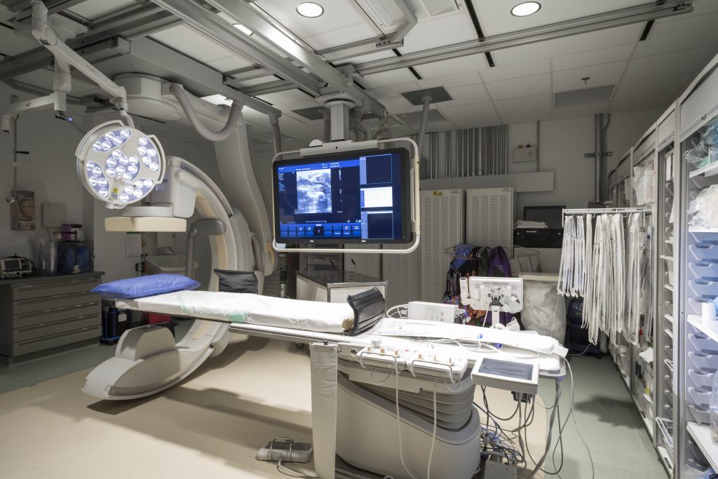
Cardiac Catheterization Lab (Cath Lab). The big C-shaped structure, is the fluoroscopy unit.

Cardiac Catheterization Lab (Cath Lab). The big C-shaped structure, is the fluoroscopy unit.

Cardiac catheterization is one of the invasive procedures performed by Dr. Rasi.  Interventional cardiology, the subspecialty of cardiology in which Dr. Rasi is particularly trained, involves additional training in performing this procedure. This procedure is also known commonly by people as stenting, angioplasty etc., but each of these terms mean very specific things which we will discuss below.  For further information, please see the separate article here about cardiac catheterization.

 In this particular segment, we will be looking primarily at the patient experience with cardiac catheterization, and how to prepare for it. The procedure of cardiac catheterization is not surgery. The only needles involved, and knows cutting suturing or open heart surgery is involved with this. In the vast majority of cases, general anesthesia is not involved either  in the procedures performed under conscious sedation.

 Cardiac catheterization is done in the hospital. Prior to arriving at the hospital, someone will call you to explain what to expect. This typically involves the following steps, so please read this information carefully.  This information is not meant to be a substitute for personalized instructions that may be given to you by the hospital. This is simply a general set of rules that apply when Dr. Rasi is performing cardiac catheterization.


 Cath Lab preprocedure checklist

  1. If you have not done so, please have your labs drawn two to three days  prior to undergoing the procedure. This will enable Dr. Rasi to ensure that your kidney function, electrolytes, and blood counts are all within normal limits before the procedure.
  2.  If you are taking any blood thinners such as warfarin (coumadin),  dabigatran (pradaxa), rivaroxaban (xarelto) or apixaban (eliquis),  please notify Dr. Rasi, or one of his medical assistants. Depending on the medication you are taking, these medications will need to be stopped approximately 3 days before the procedure,  to minimize the risk of bleeding.  This restriction does not apply to you, if you are taking aspirin and/or clopidogrel (plavix) and/or ticagrelor (brilinta).
  3.  If you have a prior documented allergy to contrast dye, please let Dr. Rasi or one of his medical assistants know.  you will need to go through a series of pre-medications in order to undergo this procedure safely.
  4.  If you have had a prior allergic reaction  to sedation medications such a Fentanyl or midazolam (versed),  please also let us know since these medications would be administered to you during the procedure.
  5.  If you have had any prior surgeries in your blood vessels of the leg, or in your aorta (vascular stents, lower extremity bypass surgery, or aortic aneurysm or aortic surgery),  please let the staff know.
  6.  If you have any restrictions that prevent us from transfusing you with blood due to religious reasons or otherwise,  please let us know.
  7.  You are expected to come to the hospital in a fasting state, after not having eaten anything for approximately 8-12 hours.

what to expect during the procedure

images.jpg

 Before the procedure begins,  the staff at the hospital will place  an IV in your arm,  and start you on some IV fluids. They will then transport you to the cardiac catheterization lab where you will be asked to rest on your back on the table. Surrounding the table, you will find a large machine with a moving arm which is the fluoroscopy unit. Think of this machine as a video x-ray machine that will show Dr. Rasi the blood vessels inside your heart so that he can both interrogate the patency of these vessels, and perform a procedure to correct it if there is any blockage.

First of all, Dr. Rasi will give you some medications through the IV to get you sedated. These medications will help you relax, and might even make you sleepy. The intent is to make you comfortable, and not to completely knock you out.  Thereafter, using an ultrasound machine, he will apply  lidocaine, a local anesthetic similar to what your dentist uses to numb up the skin near your leg and groin.  Once the femoral artery ( the major artery that runs down your leg)  has been located by ultrasound, a needle will be carefully passed into that artery, and using a very small wire, this needle would be exchanged for a tapered tube called a sheath.  The sheath is a plastic tube with a one-way valve that enables the doctor to place catheters ( small flexible plastic tubes)  into your femoral artery, without causing blood to travel in the opposite direction and come out of the body.  Once the sheath has been placed, you should have no further discomfort whatsoever.  Dr. Rasi will then begin the cardiac catheterization procedure, where small plastic tubes are advanced through the sheath and used to engage the arteries that directly supply blood to the heart called the coronary arteries.  To ensure that these arteries are open,  a special dye (contrast dye)  will be injected from the outside into the artery.  This contrast has the special property that it is visible as a black opaque substance on the x-ray camera, and therefore the flow of the contrast into the artery can be easily seen on the x-ray image.  This will tell your doctor whether there is a blockage that needs fixing or not.

 Typically this is all that cardiac catheterization involves.  Once the pictures have been taken, the catheters all come out of the body, and then the sheet is removed.  After the sheath is removed, Dr. Rasi may place a special suture or a small plug to close off the hole in the artery.  Sometimes, if the location of the  hole in the artery is not favorable, the staff in the Cath Lab will simply remove the sheath and whole pressure until the blood completely clots.

 You will then lay flat for about 2 to 3 hours, and then progressively begin ambulating in the recovery area, and if all goes well and no intervention is performed, you can go home the same day.

A coronary angiogram. The black silhouette represents the contrast dye flowing through the coronary arteries.

A coronary angiogram. The black silhouette represents the contrast dye flowing through the coronary arteries.


What if something needs to be fixed?

 If something needs to be fixed after the cardiac catheterization is complete,  Dr. Rasi will notify you on whether something needs to be fixed, and whether it can be done safely.  If the procedure can be completed safely, he will more than likely place a stent or perform a balloon angioplasty to correct the lesion. If the lesion is more complex, he may then  decide to transfer you to another hospital where higher level of care including on-site cardiac surgeon might be available. Typical locations for this include the Texas Medical Center where very complicated procedures can be performed in relative safety. 

 If Dr. Rasi does decide to perform an angioplasty or place a stent, this procedure will possibly add another 45 minutes to an hour to the overall procedure time. In addition, you will receive blood thinning medication (Heparin)  to help with this process.  If a balloon angioplasty or stent placement is performed, the recovery time is longer, and you will be asked to stay overnight at the very least in the hospital. This is for the sole purposes of careful monitoring.


 For more information regarding this procedure, including some of the ways Dr. Rasi makes decisions about what to do, please see the separate section on cardiac catheterization.


pheripheral angiography : what to expect

Peripheral angiography is very similar to cardiac catheterization.  It is performed in the cardiac catheterization lab in the hospital.  Because the procedures are so similar, please refer to the section on cardiac catheterization for further information. That being said, there are important differences between the peripheral angiogram and a cardiac catheterization. The most important difference is that peripheral angiography focuses on assessing the patency and integrity of blood vessels in the lower extremities compared to blood vessels that supply blood to the heart (coronary arteries).

 From the patient standpoint, the experience is very similar. The same precautions one would take during cardiac catheterization are also undertaken during peripheral angiography. In fact,  the place where the physician will enter the blood vessels of the leg, is exactly the same as they would when cardiac catheterization is performed. There are two main differences in the way  Dr. Rasi will be performing the  peripheral angiogram.

  1.  The first difference, is that the site where the needle entry into the femoral artery happens, in the opposite leg usually, from where the blockage is suspected. This can be a confusing point for many patients. For example, if there is a nonhealing ulcer in your right leg, Dr. Rasi will place the catheter into the left leg. This discrepancy is because usually the best way to get to the left leg is from what cardiologists generally called the " up and over"  approach. 
  2.  the second difference is that the catheters never travel up the aorta into the heart. The almost always travel down the opposite leg, or down the same leg.

 The main objective in a peripheral angiogram is to place the catheter tip at the beginning of the artery that supplies blood to your leg, and injure contrast to visualize whether or not the arteries of the legs are open. If there is a blockage, depending on whether or not it is correctable, Dr. Rasi may offer one of three options.

A sample peripheral angiogram showing the blood flow through the thigh (A), knee (B) and the foot (C). 

A sample peripheral angiogram showing the blood flow through the thigh (A), knee (B) and the foot (C). 

  1.  the first option is to correct the blockage  with the use of a balloon angioplasty device, or rarely via stent placement.  This is typically the most straightforward approach.  If there is a problem accessing the vessel,  or if the blockage looks like it's more serious  Dr. Rasi may utilize a specialized device called an atherectomy device.  This  specialize device acts like the combination  of  sanding machine, along with a diamond coated drill bit. It is designed to be able to  bore through hard calcium in the peripheral arteries and facilitate the expansion of a balloon.
  2.  Sometimes,  if the blockages in the leg are too difficult to fix, Dr. Rasi may refer you to a vascular surgeon  to undergo femoral bypass surgery. 
  3.  Finally,  there may be rare options  where the only available therapy is medical therapy. This means you will be given medications in the hopes of mitigating the symptoms where no feasible mechanism exists to fix the lesion. 

 At the end of the powerful angiogram procedure, the recovery process is very similar to a cardiac catheterization.  You will be taken to the recovery area,  the sheath in your groin will be removed with the placement of a specialized closing device, or Dr. Rasi may elect to have the sheath manually removed and have someone hold pressure at that location until the vessel. You will usually spend a few hours in the recovery area and in 90% of the cases the patient go home the very same day whether or not an intervention was performed.  After every case, Dr. Rasi will discuss with you and your family the results of the procedure, and what exactly he performed.